Bactofilin / BacA protein
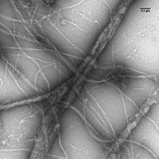
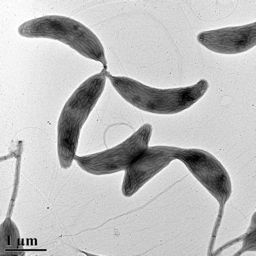
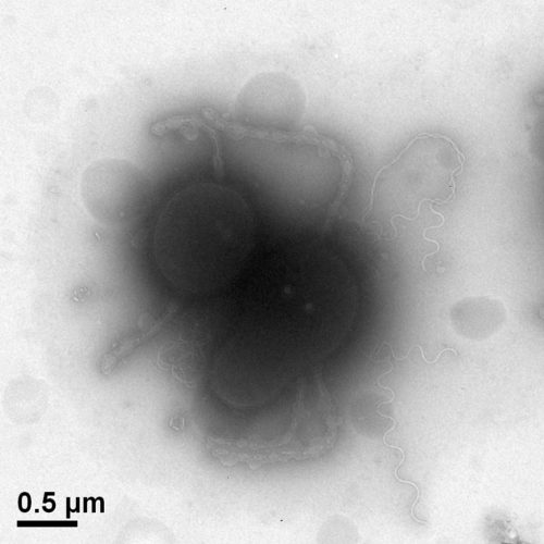
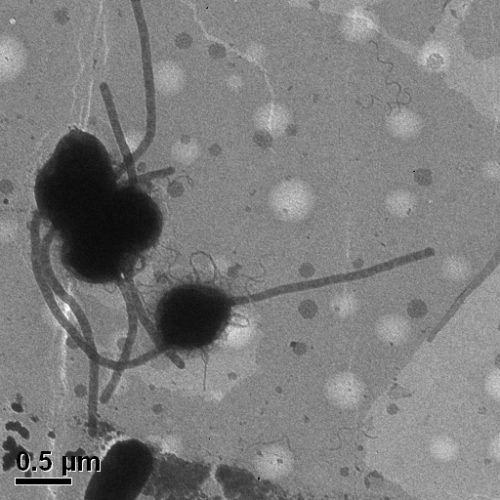
Transmission electron micrograph of Caulobacter (Berne)
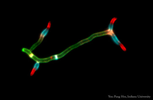
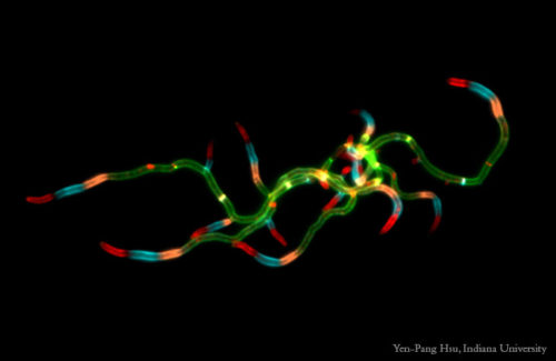
S. venezuelae FDAA (Hsu)
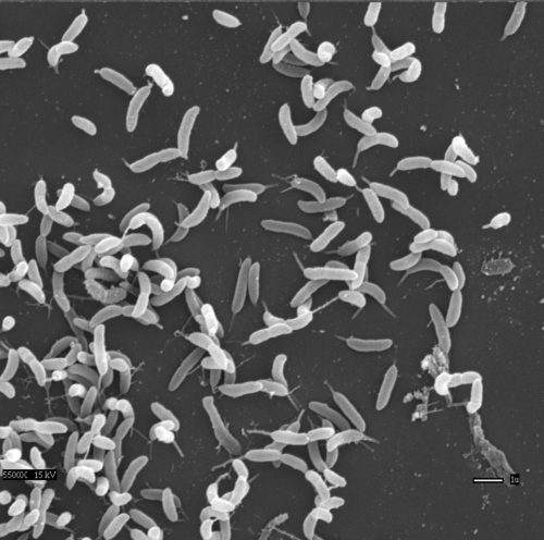
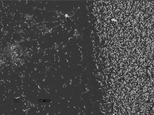
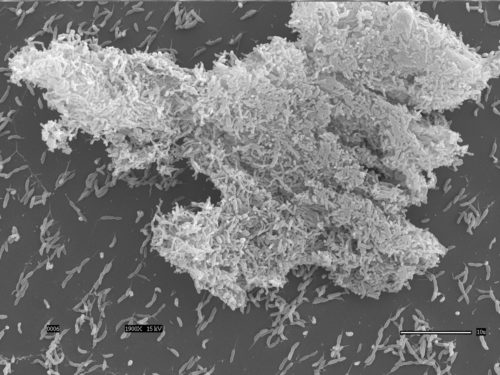
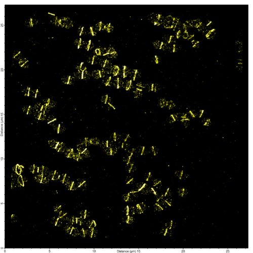
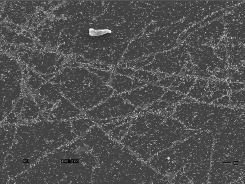
Early stage biofilm of Caulobacter attached to glass coverslip viewed with scanning electron microscope, scale bar=1µm (Berne)
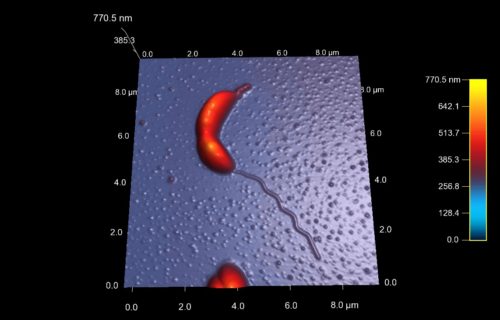
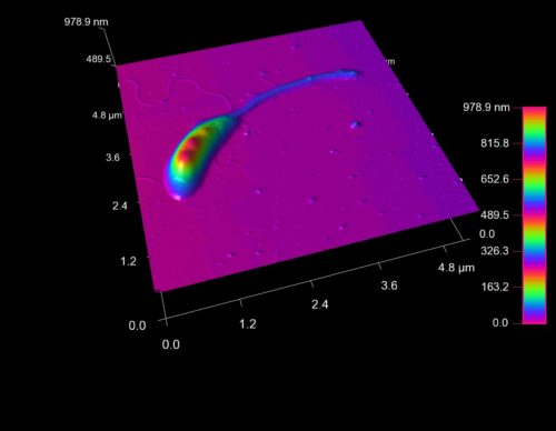
Dividing Caulobacter viewed with atomic force microscopy (Berne)
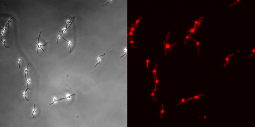
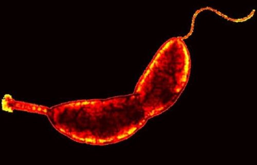
Asticcacaulis biprosthecum cells, membrane=red (Jacq)
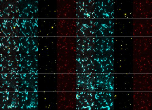
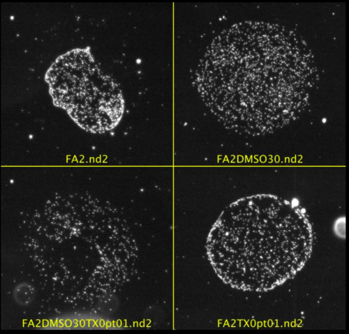
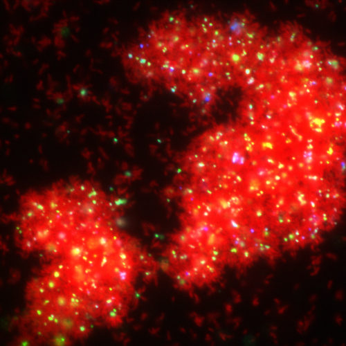
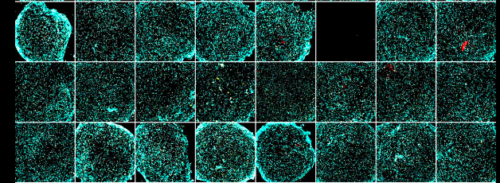
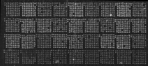
Bacterial microarray images of multiple Caulobacter mutants expressing cytoplasmic CFP, IbpA-YFP, and mCherry-SpmX (Kysela)
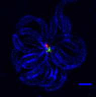
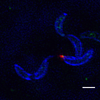
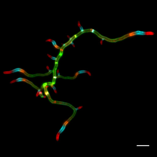
Caulobacter rosette viewed with structural illumination super-resolution microscopy: cell wall=blue, holdfast=red, DNA=green (Berne)
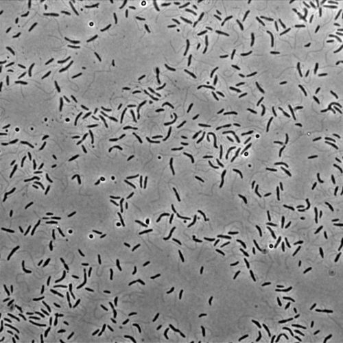
Caulobacter crescentus (Brun)
Clonal cultures of Caulobacter in microarray with different buffers (Kysela)
Caulobacter biofilm gradient (Berne)
Transmission electron micrograph of Caulobacter cresentus (Berne)
Mature biofilm of Caulobacter attached to glass coverslip viewed with scanning electron microscope, scale bar=10µm (Berne)
S. venezuelae FDAA (Hsu)
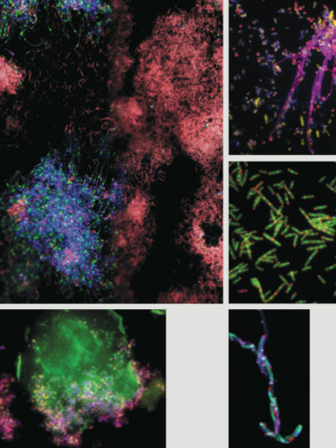
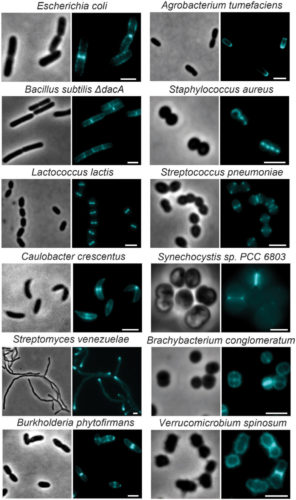
HADA labeling in various species (Hsu)
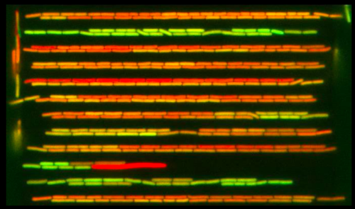
Fluorescence image of thousands of Caulobacter crescentus cells in a biofilm (Berne)
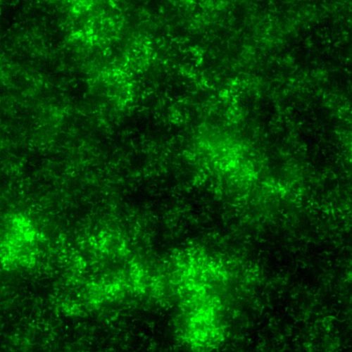
Confocal micrograph of mature Caulobacter biofilm expressing cytoplasmic green fluorescent protein (Berne)
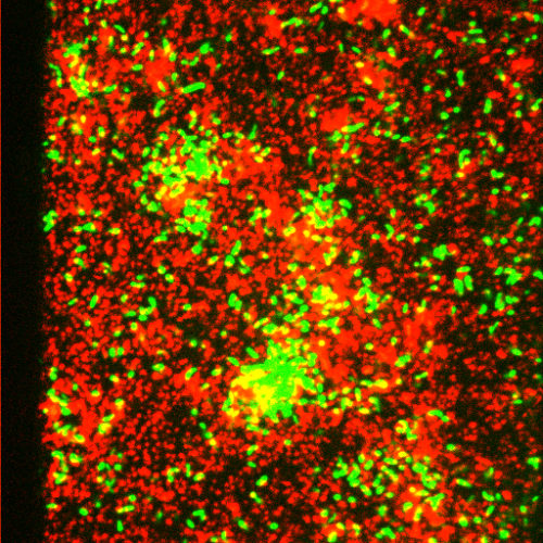
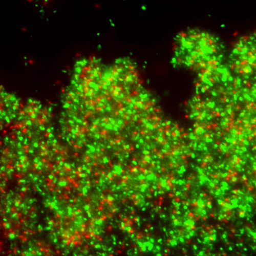
Mature Caulobacter biofilm at 72 hours of wildtype=red and pilus mutant=green (Berne)
Streptomyces venezuelae Rotor FDAA (Hsu)
PALM (Jacq)
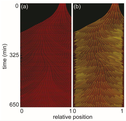
Timelapse of Caulobacter cells growing in nanochannels. Cells expressing cytoplasmic DsRed and IbpA-YFP (Kysela)
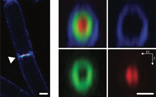
Caulobacter viewed with structural illumination super-resolution microscopy: cell wall=blue, holdfast=red (Berne)
Mature Caulobacter biofilm showing live=green and dead=red cells (Berne)
Asticcacaulis ac460 (Randich)
Phenylobacterium conjunctum
B.subtilis Virtual time-lapse labeling division sites (Hsu)
Streptomyces venezuelae FDAA (Hsu)
Parvularcula lutaonensis-TEM (Randich)
Bacillus subtilis growing in nanochannel device (cytoplasm=red, motility genes=green) (Kysela).
Streptomyces venezuelae Rotor FDAA (Hsu)
Caulobacter microarray experiment on centering image (Kysela)
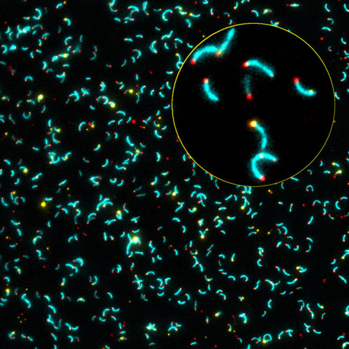
Caulobacter cells=blue, inclusion bodies=yellow, SpmX=red (Kysela)
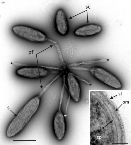
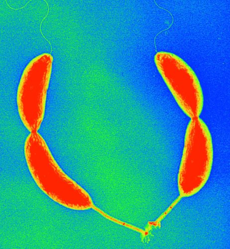
Two asymmetric predivisional cells of Caulobacter attached by their holdfasts
Darkfield microscopy image of bacterial microarray (Kysela)
Speed 50X, DNA=red, Vibrio cell=green
Chlamydia FtsZ Ring (Hsu)
Caulobacter crescentus cell with long stalk viewed with atomic force microscopy (Berne)
Bacillus subtilis growth in one nanochannel. Cytoplasm expressing red fluorescent mCherry, lineage tracked in yellow. (Kysela)
Parvularcula lutaonensis TEM (Randich)
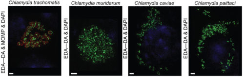
Chamydia PG staining
Caulobacter in lines on glass cover slip, scanning electron micrograph(Berne)
PG ring in intracellular Chlamydia cells (Hsu)
Videos